 |
|
|
|
LSY-30036B Bovine Brucellosis Indirect ELISA Antibody Test Kit Brucella Abortus assay
Product Details:
Payment & Shipping Terms:
|

Detailed Product Description
| Sample Performance: | Serum Of Bovine | Shelf Life: | 12 Months When Store At 2~8℃ |
|---|---|---|---|
| Incubation Temperature: | 37℃ | Incubation Time: | 30min-30min-10min |
Bovine Brucellosis Indirect ELISA Antibody Test Kit
Catalog No.: LSY-30036B
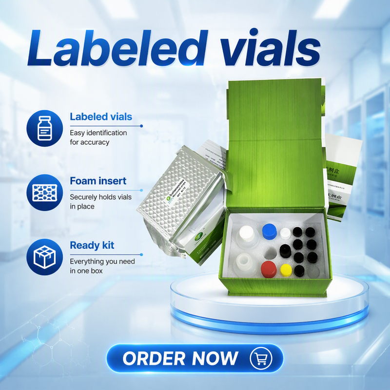
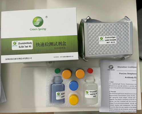
[ Components and Contents]
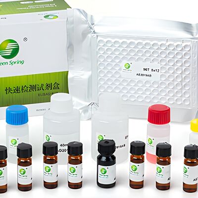
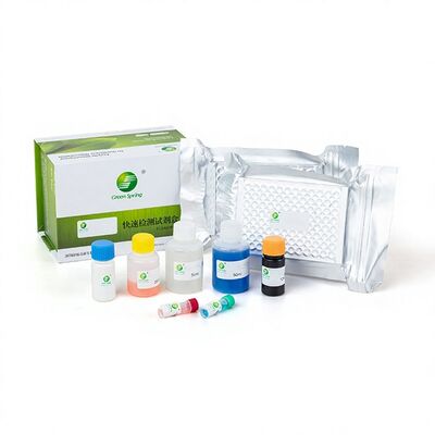
| Code | Kit Components | Quantity |
| 1 | Brucellosis antigen-coated plate | 96 wells/plate × 2 plates |
| 2 | Sample diluent | 50 ml/bottle × 1 bottle |
| 3 | Negative control | 1.5 ml/ vial × 1 vial |
| 4 | Positive control | 1.5 ml/ vial × 1 vial |
| 5 | 10×concentrated washing buffer | 50 ml/bottle × 1 bottle |
| 6 | Enzyme conjugate | 22 ml/bottle × 1 bottle |
| 7 | Substrate | 12 ml/bottle × 2 bottles |
| 8 | Stop solution | 12 ml/bottle × 1 bottle |
| 9 | Adhesive Foil | 2 sheets |
| 10 | Instructions | 1 copy |
[Function and Purpose]
Brucellosis is a zoonotic infectious disease caused by Brucella bacteria. It is characterized by inflammation, necrosis, and the formation of granulomas in the reproductive organs, fetal membranes, and various other organ tissues, resulting in symptoms such as miscarriage, infertility, orchitis, and arthritis. A variety of animals exhibit varying degrees of susceptibility to the disease; natural infections are most common in sheep, cattle, and pigs. Animal infection can occur through multiple routes, including the digestive tract, the skin and mucous membranes, and vertical transmission. Brucellosis poses a serious threat to human health and adversely affects the development of animal husbandry in the worldwide.
The microwell plates in this kit are coated with purified Brucella antigens; the indirect ELISA method is used to detect Brucella antibodies in bovine serum. The control and the diluted serum sample are added to the wells; if Brucella antibodies are present in the sample, an antigen–antibody complex will form. After washing the plate, horseradish peroxidase (HRP)-labeled antibodies are added to the wells; the enzyme conjugate binds to the Brucella antibodies, forming an "antigen–antibody–enzyme conjugate" complex. After washing the plate to remove excess enzyme conjugate, a substrate solution (TMB) is added, which is enzymatically catalyzed to produce a blue product. The color development depends on the amount of specific antibodies present in the test sample: if antibodies are present, a blue color develops; after the addition of the stop solution, the color changes to yellow; if no antibodies are present, no color reaction occurs. The absorbance (OD) of the reaction plate is measured using a microplate reader at a wavelength of 450 nm.
[Usage and Determination]
1 Preparation before use
1.1 Sample Preparation: Collect whole animal blood; after the blood has coagulated, allow the serum to separate naturally, or centrifuge at 4000 rpm/min for 10 minutes and collect the supernatant. The serum should be clear and free of hemolysis.
1.2 Experimental Preparation: All reagents in the kit must be returned to room temperature (18–26℃) before use; shake thoroughly before adding the solution.
1.3 Preparation of Washing buffer: Dilute the 10×concentrated washing buffer with deionized water at 10 times. (10ml 10×concentrated washing buffer + 90ml deionized water ) It is normal if there is crystallization in the
10×concentrated washing buffer, put at 37℃ water bath until completely dissolved.
1.4 Sample Dilution: Dilute the serum sample with the sample diluent for 40times (for example, add 5μl of serum to 195μl of sample diluent), and mix thoroughly.
Note: Negative controls and positive controls do not require dilution.
2 Operating Steps
2.1 Add Sample: Take an antigen-coated plate (the plate may be opened and used in separate portions according to sample quantity); add 100 µl of the diluted sample to each corresponding well, meanwhile set 2 wells for positive control, 2 wells for negative control , add positive control or negative control 100µl/well to it’s well accordingly.
2.2 Incubation: After adding the sample, seal the plate with the Adhesive Foil and incubate at 37℃ for 30 minutes.
2.3 Plate washing: remove the liquid from the wells, add 300μl of washing buffer per well, and let stand for 10 seconds; remove the liquid again, wash three times, drying the plate with absorbent paper during the final wash.
2.4 Add Enzyme conjugate: Add 100 µl of Enzyme conjugate to each well.
2.5 Incubation: Cover with the Adhesive Foil and incubate at 37℃ for 30 minutes.
2.6 Plate washing: follow step 2.3.
2.7 Add substrate: add 100 µl of substrate to each well, gently shake to mix, and incubate at 37℃ in the dark for 10 minutes.
2.8 Stop reaction: Add 50 µl of stop solution to each well, gently shake for 10 seconds, mix thoroughly, and read the absorbance immediately after termination.
2.9 Reading: measure at a wavelength of 450nm and record the OD value for each well by ELISA reader.
3. Result judgement
3.1 The average OD value of the positive control is designated as ODPC; the average OD value of the negative control is designated as ODNC; and the OD value of the sample is designated as ODS.
3.2 For the assay to be valid: The ODPC value≥0.60 and the ODNC value <0.20; Otherwise, the experiment must be repeated.
3.3 Calculate the S/P value= (ODS–ODNC) / (ODPC–ODNC).
3.4 Determination: Under the condition that the assay is valid, an S/P value ≥ 0.20 is considered positive; an S/P value <0.20 is considered negative.
Shenzhen Lvshiyuan Biotechnology Co., Ltd
D Building, National Biological Industrial Park of Marinelife, No.2 Binhai Road, Dapeng, Shenzhen, 518120 China
Tel. 86-755-28438788
Fax 86-755-28938800
Email: info@lsybt.com
www.lsybt.com

Contact Details
Other Products
-
Canine/ Feline/ Rabbit/ Rat Toxoplasma gondii IgG antibody ELISA kit TOXO Ab
-
Foot and mouth disease virus (FMDV) Type O Antibody Test (block ELISA) animal diagnostic kits
-
Gumboro Disease (IBD) Virus Antibody test kit infectious bursal disease elisa
-
Infectious Bronchitis(IB) IgG antibody diagnostics kit poultry elisa test
-
Avian Marek's disease virus antibody ELISA test veterinary diagnostic kit for mareks
-
Avian EDS76 antibody ELISA kit egg drop syndrome detection